
ꯐꯥꯏꯜ:Animal Cell.svg

Size of this PNG preview of this SVG file: ৮০০ × ৪৬২ ꯄꯤꯛꯆꯦꯜꯁ. ꯑꯇꯩ ꯁꯦꯡꯅ ꯌꯦꯡꯕ ꯌꯥꯕ: ৩২০ × ১৮৫ ꯄꯤꯛꯆꯦꯜꯁ | ৬৪০ × ৩৬৯ ꯄꯤꯛꯆꯦꯜꯁ | ১,০২৪ × ৫৯১ ꯄꯤꯛꯆꯦꯜꯁ | ১,২৮০ × ৭৩৯ ꯄꯤꯛꯆꯦꯜꯁ | ২,৫৬০ × ১,৪৭৮ ꯄꯤꯛꯆꯦꯜꯁ | ১,৪০৫ × ৮১১ ꯄꯤꯛꯆꯦꯜꯁ.
ꯐꯥꯏꯜ ꯑꯁꯦꯡꯕ (ꯑꯦꯁ•ꯚꯤ•ꯖꯤ ꯐꯥꯏꯜ, ꯌꯥꯝꯗꯅ ১,৪০৫ × ৮১১ ꯄꯤꯛꯆꯦꯜ, ꯐꯥꯏꯜ ꯆꯥꯎꯕꯒꯤ ꯆꯥꯡ: ৪৫৭ KB)
ꯑꯇꯦꯟꯕꯥ ꯁꯟꯗꯣꯛꯅꯥ ꯇꯥꯛꯄꯥ
| ꯁꯟꯗꯣꯛꯅꯥ ꯇꯥꯛꯄꯥ |
English: A reworked version of File:Biological_cell.svg.
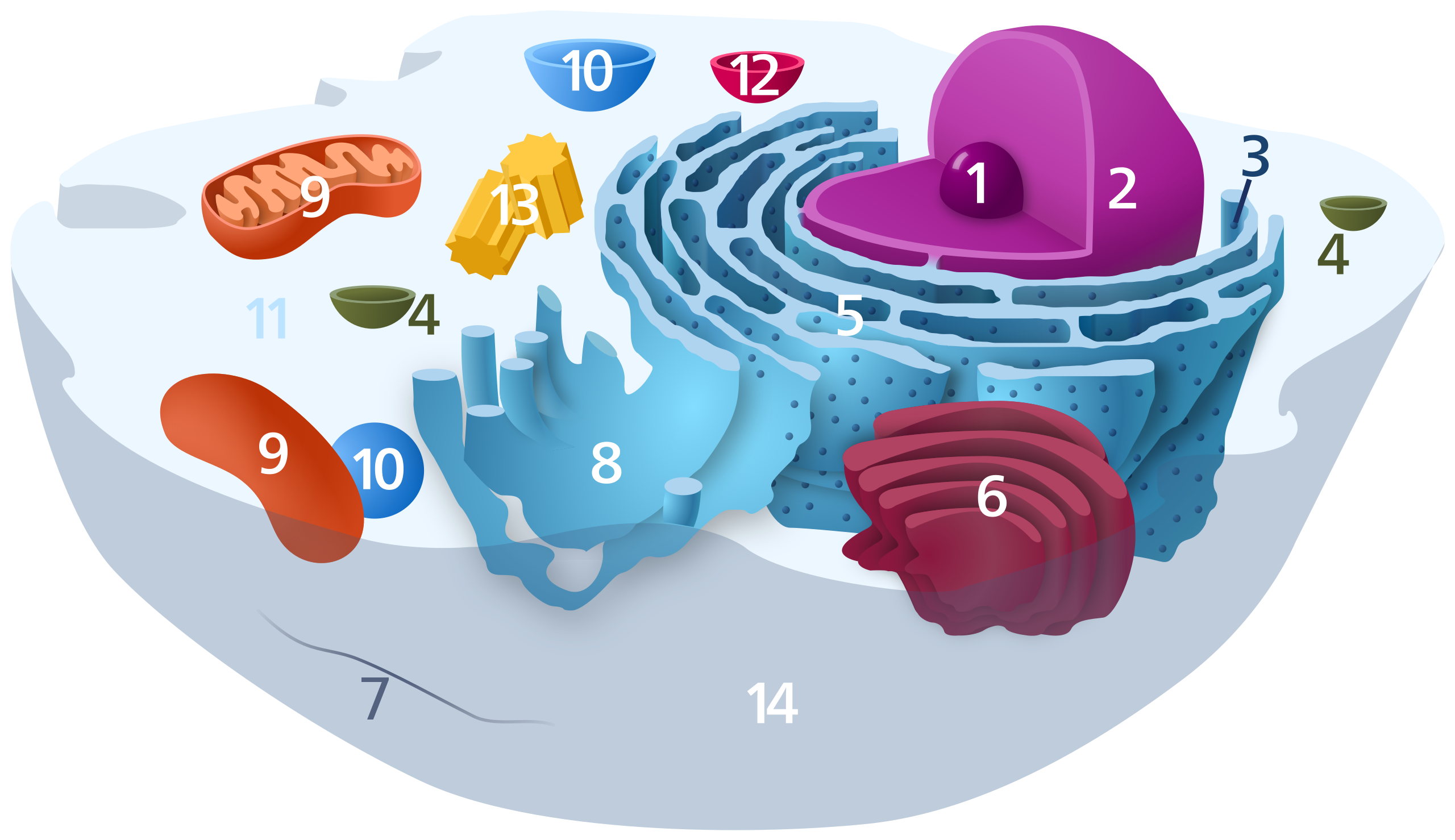
Diagram of a typical animal cell. Organelles are labelled as follows:
العربية: رسم تخطيطي للخلية الحيوانية
Català: Dibuix esquemàtic d'una cèl·lula animal típica:
Español: Diagrama de una célula animal típica:
ਪੰਜਾਬੀ: ਕਿਸੇ ਮਿਸਾਲੀ ਜਾਨਵਰ ਦੇ ਕੋਸ਼ਾਣੂ ਦਾ ਚਿੱਤਰ:
Svenska: Schematisk bild över en typisk eukaryot cell, som visar cellens subcellulära komponenter. Organeller:
Deutsch: Organisation einer typischen eukaryotischen Tierzelle:
|
|||
| ꯆꯩꯆꯠ | ||||
| ꯍꯧꯔꯛꯐꯛ | ꯏꯁꯥ ꯏꯊꯟꯇꯒꯤ ꯑꯣꯏꯕꯥ ꯊꯕꯛ | |||
| ꯄꯨꯊꯣꯛꯂꯤꯕ ꯃꯤ | Kelvinsong | |||
| ꯑꯌꯥꯕ ꯂꯧꯕ (ꯃꯁꯤꯒꯤ ꯐꯥꯏꯜꯁꯤ ꯑꯃꯨꯛ ꯍꯟꯅꯥ ꯁꯤꯖꯤꯟꯅꯕꯥ) |
I, the copyright holder of this work, hereby publish it under the following license:
|
ꯐꯥꯏꯜꯒꯤ ꯄꯨꯋꯥꯔꯤ
ꯆꯩꯆꯠ/ꯃꯇꯝꯗ ꯅꯝꯃꯨ ꯃꯇꯝ ꯑꯗꯨꯗ ꯐꯥꯏꯜ ꯑꯗꯨ ꯎꯅꯕ
| ꯆꯩꯆꯠ/ꯃꯇꯝ | ꯈꯨꯠꯄꯤꯈꯨꯖꯤꯟ | ꯄꯥꯛ ꯆꯥꯎꯕꯥ | ꯁꯤꯖꯤꯟꯅꯔꯤꯕ | ꯑꯄꯥꯝꯕ ꯐꯣꯡꯗꯣꯛ ꯎ | |
|---|---|---|---|---|---|
| ꯍꯧꯖꯤꯛꯀꯤ | ꯲꯰:꯱꯷, ꯱꯷ ꯅꯣꯚꯦꯝꯕꯔ ꯲꯰꯲꯲ | | ১,৪০৫ × ৮১১ (৪৫৭ KB) | TheBartgry | Reverted to version as of 00:21, 10 December 2012 (UTC) showing continuity between nuclear membrane and ER is useful |
| ꯰꯷:꯰꯲, ꯲꯶ ꯖꯨꯂꯥꯏ ꯲꯰꯲꯱ |  | ১,৪০৫ × ৮১১ (৪৫২ KB) | FabPon | Reverted to version as of 00:17, 2 December 2012 (UTC) | |
| ꯰꯵:꯵꯱, ꯱꯰ ꯗꯤꯁꯦꯝꯕꯔ ꯲꯰꯱꯲ |  | ১,৪০৫ × ৮১১ (৪৫৭ KB) | IsadoraofIbiza | Showing Nuclear membrane—ER continuity | |
| ꯰꯵:꯴꯷, ꯲ ꯗꯤꯁꯦꯝꯕꯔ ꯲꯰꯱꯲ |  | ১,৪০৫ × ৮১১ (৪৫২ KB) | IsadoraofIbiza | center | |
| ꯰꯵:꯳꯷, ꯲ ꯗꯤꯁꯦꯝꯕꯔ ꯲꯰꯱꯲ |  | ১,৪৬৬ × ৮৯১ (৪৫৫ KB) | IsadoraofIbiza | Add cytoskeleton | |
| ꯰꯵:꯳꯳, ꯲ ꯗꯤꯁꯦꯝꯕꯔ ꯲꯰꯱꯲ |  | ১,৪৬৬ × ৮৯১ (৪৫৩ KB) | IsadoraofIbiza | User created page with UploadWizard |
ꯐꯥꯏꯜꯒꯤ ꯁꯤꯖꯤꯟꯅꯐꯝ
ꯃꯇꯨꯡ ꯏꯟꯕ ꯂꯃꯥꯏꯁꯤꯖꯤꯟꯅꯕ ꯃꯁꯤꯒꯤ ꯐꯥꯏꯜ:
ꯃꯥꯂꯦꯝꯒꯤ ꯊꯥꯛꯇꯥ ꯁꯤꯖꯤꯟꯅꯕꯥ ꯐꯥꯏꯜ
ꯃꯁꯤꯒꯤ ꯐꯥꯏꯜ ꯑꯁꯤ ꯃꯈꯥꯒꯤ ꯑꯇꯩ ꯋꯤꯀꯤꯁꯤꯡꯅ ꯁꯤꯖꯤꯟꯅꯩ:
- an.wikipedia.org ꯗꯥ ꯁꯤꯖꯤꯟꯅꯩ
- ar.wikipedia.org ꯗꯥ ꯁꯤꯖꯤꯟꯅꯩ
- جهاز غولجي
- ميتوكندريون
- جسيم حال
- نواة (خلية)
- ريبوسوم
- عضية خلوية
- بوابة:علم الأحياء
- هيكل خلوي
- بوابة:علم الحيوان
- بوابة:علم الأحياء/بوابات شقيقة
- شبكة إندوبلازمية
- علم الخلية
- جسم بلعمي
- نوية (خلية)
- نظام غشائي داخلي
- سيتوبلازم
- بوابة:علم الحيوان/بوابات شقيقة
- فجوة عصارية
- جسيم مركزي
- بوابة:سنوريات
- قالب:مخطط العضيات
- بوابة:سنوريات/بوابات شقيقة
- جسيم حال بلعمي
- عصارة خلوية
- قالب:مخطط العضيات/عرضي
- bn.wikipedia.org ꯗꯥ ꯁꯤꯖꯤꯟꯅꯩ
- br.wikipedia.org ꯗꯥ ꯁꯤꯖꯤꯟꯅꯩ
- bs.wikipedia.org ꯗꯥ ꯁꯤꯖꯤꯟꯅꯩ
- ca.wikipedia.org ꯗꯥ ꯁꯤꯖꯤꯟꯅꯩ
- ckb.wikipedia.org ꯗꯥ ꯁꯤꯖꯤꯟꯅꯩ
- da.wikipedia.org ꯗꯥ ꯁꯤꯖꯤꯟꯅꯩ
- de.wikipedia.org ꯗꯥ ꯁꯤꯖꯤꯟꯅꯩ
ꯌꯦꯡꯉꯨ ꯃꯂꯦꯝꯒꯤ ꯊꯥꯛꯀꯤ ꯁꯤꯖꯤꯟꯅꯐꯝ ꯑꯗꯨ ꯃꯁꯤꯒꯤ ꯐꯥꯏꯜꯁꯤꯗ ꯫